38 provide the labels for the electron micrograph
Electron Micrographs of Cell Organelles - Biology Discussion It is an electron micrograph of cell's largest and most important organelle - the mitochondria and is characterized by the following features (Fig. 7 & 8): (1) The name mitochondria was given by Benda (1898) and their ma n function was brought to light by Kingsbury (1912). (2) Each mitochondria in section appears as sausage or cup or bowl ... histology.medicine.umich.edu › resources › renal-systemRenal System | histology Correct answer 4. A podocyte - Note that you can see areas where the cytoplasm of the cell is connected to the pedicels (foot processes, labeled "p"). "V" labels vascular lumen. "E" indicates endothelial cell cytoplasm. "f" indicates an endothelial fenestra. The bracket indicates basal lamina. "U" indicates urinary space.
Specialty Expiration Labels, Electron Microscopy Sciences 100489-622PK 36.72 USD. 100489-622. Specialty Expiration Labels, Electron Microscopy Sciences. Labels. Quickly identify outdated laboratory items such as reagents and controls. These labels provide space for noting the date that the items were received, opened and when it will expire. A permanent adhesive holds the label firmly in place on a ...

Provide the labels for the electron micrograph
Identify The Bands And Lines Of The Striations Of A Tem Of Relaxed ... Figure 18.5 Identify the bands and lines of the striations in this transmission electron micrograph of relaxed sarcomeres (8,400x). Part C Complete the following statements: _of a muscle is usually attached to a fixed part. _of a muscle is usually attached to a movable part. 3. The forearm is flexed at the elbow when the_muscle contracts. 4. Integumentary System | histology 1. Letter A labels the reticular dermis, so named because of the network of coarse, type I collagen fibers (Letter B indicates the papillary dermis). 2. Letter C labels Meissner's corpuscles, which are mechanosensory receptors that respond primarily to light touch and low frequency stimuli. 3. Micrograph - an overview | ScienceDirect Topics The SEM micrograph of S41, shown in Fig. 5a, reveals the presence of regular spheres with diameters in the range between 500 - 1000 nm. For C41 (Fig. 5b) a sponge like structure with pores > 1 μm is observed. The micrograph of "as made" Z41(50) (Fig. 5c) shows roughly cubic crystals with rounded edges and a size in the range 500 - 800 nm.
Provide the labels for the electron micrograph. Hydroponic Lettuce Was Seen as Safe From Salmonella, Until an … May 03, 2022 · A colored scanning electron micrograph of Salmonella typhimurium. The strain of bacteria sickened 31 people last summer, and was found in a storm-water basin near the BrightFarms facility. Credit... Virtual EM Micrograph List | histology 021. Plasma Cell: This electron micrograph shows a typical secretory cell, a plasma cell, which secretes immunoglobulin protein. Many of the major types of cellular organelles are visible in this image. In the nucleus, areas of euchromatin and heterochromatin can easily be identified. Virtual Slide. Label This Transmission Electron Micrograph : TEM of chloroplast from ... Label the transmission electron micrograph of the nucleus. Provide the labels for the electron micrograph in figure 12.8. Labeling for electron microscopy using antibody conjugated to. Transmission electron microscopy (tem) is a microscopy technique in which a beam of electrons is transmitted through a specimen to form an image. What is Electron Microscopy? - UMASS Medical School Electron microscopy is used in conjunction with a variety of ancillary techniques (e.g. thin sectioning, immuno-labeling, negative staining) to answer specific questions. EM images provide key information on the structural basis of cell function and of cell disease.
Provide the labels for the electron micrograph in figure 1 ... - Chegg Problem 3PAB Provide the labels for the electron micrograph in figure 19.5. FIGURE 19.5 Identify the bands and lines of the striations of a transmission electron micrograph of relaxed sarcomeres (16,000×), using the terms provided. ________________________________________________________ Step-by-step solution This problem hasn't been solved yet! › books › NBK26880Looking at the Structure of Cells in the Microscope Moreover, in recent years, the performance of electron microscopes has been improved by the development of electron illumination sources called field emission guns. These very bright and coherent sources can substantially improve the resolution achieved. The major landmarks in the development of electron microscopy are listed in Table 9-2. Solved: Chapter 20 Problem 1LAB Solution - Chegg Access Laboratory Manual for Human Anatomy & Physiology Main Version 3rd Edition Chapter 20 Problem 1LAB solution now. Our solutions are written by Chegg experts so you can be assured of the highest quality! Identifying long-range synaptic inputs using genetically encoded labels ... Enzymes that facilitate the local deposition of electron dense reaction products have been widely used as labels in electron microscopy (EM) for the identification of synaptic contacts in neural ...
Renal System | histology Correct answer 4. A podocyte - Note that you can see areas where the cytoplasm of the cell is connected to the pedicels (foot processes, labeled "p"). "V" labels vascular lumen. "E" indicates endothelial cell cytoplasm. "f" indicates an endothelial fenestra. The bracket indicates basal lamina. "U" indicates urinary space. Looking at the Structure of Cells in the Microscope A typical animal cell is 10–20 μm in diameter, which is about one-fifth the size of the smallest particle visible to the naked eye. It was not until good light microscopes became available in the early part of the nineteenth century that all plant and animal tissues were discovered to be aggregates of individual cells. This discovery, proposed as the cell doctrine by Schleiden and … Muscle Lab 19 Figure 19.5 Sarcomere Diagram - Quizlet THagge TEACHER. Chapter 5 - Tissues Lecture Exam. 61 Terms. THagge TEACHER. Lab 14: Figure 14.10 Anterior Features of the Skull. 9 Terms. THagge TEACHER. Lab 14: Figure 14.11 Lateral View of the Skull. 14 Terms. 8 Human AP - Course Hero Question Provide the labels for the electron micrograph in figure 19.5 Image transcription text 8 Human AP | Lab Terms: A band (dark) emps I band (light) Sarcomere 2 Z line H zone M line 3 GURE 19.5 Using the terms provided, identify the bands and lines of the striations of a transmission electron crograph of relaxed sarcomeres... Show more ...
Scanning electron microscopy showing the microstructure of collagen ...
PDF Electron Micrographs (EMs) for laboratories in A215, Basic Human Anatomy There are distinct differences between cilia and microvilli to be seen in electron micrographs: - Cilia are larger (the cilium labeled C about 2.5 microns along its length is probably 5 to 10 microns long); - Cilia contain microtubules, by which they can move.
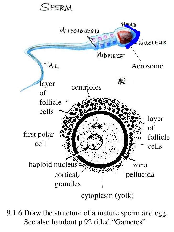
IB Biology Draw assessment statements
animal cell electron micrograph labelling Diagram - Quizlet Start studying animal cell electron micrograph labelling. Learn vocabulary, terms, and more with flashcards, games, and other study tools.

Lysosomes Dr.Jastrow's electron microscopic atlas
The Mitochondrion - Molecular Biology of the Cell - NCBI … The process of electron transport begins when the hydride ion is removed from NADH (to regenerate NAD +) and is converted into a proton and two electrons (H-→ H + + 2e-). The two electrons are passed to the first of the more than 15 different electron carriers in the respiratory chain. The electrons start with very high energy and gradually ...
34 Label The Transmission Electron Micrograph Of The Nucleus - Labels 2021
Labeling the Cell Flashcards | Quizlet Label the transmission electron micrograph of the nucleus. membrane bound organelles golgi apparatus, mitochondrion, lysosome, peroxisome, rough endoplasmic reticulum nonmembrane bound organelles ribosomes, centrosome, proteasomes cytoskeleton includes microfilaments, intermediate filaments, microtubules Identify the highlighted structures

| ER bodies in Arabidopsis thaliana. A confocal micrograph (A) and an ...
PDF Identifying Organelles from an Electron Micrograph Courtesy of Dr. Julian Thorpe - EM & FACS Lab, Biological Sciences University Of Sussex The electron micrograph displayed below illustrates many of the plant cell characteristics discussed The cell wall, large central vacuole and chloroplasts are clearly visible Also visible is the clearly defined nucleus containing chromatin

(PDF) In Situ Study of the Impact of Aberration-Corrected Electron-Beam ...
Diagram of labeling used for light and electron microscopy. A: Labeling ... An ideal label for light and electron microscopy would contain both fluorescent dye and a colloidal nanoparticle on a single antibody molecule. Secondary and tertiary label, the colloidal ...

Schwann cells Dr.Jastrow's electron microscopic atlas
Solved: Chapter 20 Problem 1LAB Solution - Chegg Access Laboratory Manual for Human Anatomy & Physiology 2nd Edition Chapter 20 Problem 1LAB solution now. Our solutions are written by Chegg experts so you can be assured of the highest quality!

Animal Cell - Electron Microscope - Worksheet | Animal cell structure ...
Researchers uncover a novel host cell pathway hijacked during … Jun 14, 2022 · An international team of scientists, led by the University of Bristol, has been investigating how the SARS-CoV-2 virus, the coronavirus responsible for the COVID-19 pandemic, manipulates host ...

Transmission electron microscopy of cysts produced in cell culture. (A ...
medicalxpress.com › news › 2022-06-uncover-host-cellResearchers uncover a novel host cell pathway hijacked during ... Jun 14, 2022 · An international team of scientists, led by the University of Bristol, has been investigating how the SARS-CoV-2 virus, the coronavirus responsible for the COVID-19 pandemic, manipulates host ...
Post a Comment for "38 provide the labels for the electron micrograph"